




The Reality of Endodontic Flare-Ups: Definitions, Incidence, Key Risk Factors, and Clinical Management
After root canal treatment, some patients experience unexpected severe pain or swelling. This is commonly called a “flare-up,” and its incidence, based on a strict definition, has been reported to be about 2.83%. While it occurs in roughly 3 out of 100 patients, several factors overlap in the background, including preoperative infection and pain status as well as fine procedural considerations. In this article, we organize, in a clinic-friendly way, why flare-ups occur and key points for their prevention and management, based on the latest evidence.
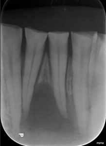
The first point to understand is “who is more likely to experience it.” The clearest sign is preoperative pain. In particular, when there is spontaneous pain (throbbing without stimulation) or tenderness to percussion (pain on biting or tapping), acute postoperative symptoms are more likely. Evidence suggests that spontaneous pain increases relative risk by about 5.8 times and percussion pain by about 3.5 times. In addition, cases showing a radiolucency at the apex on radiographs (apical periodontitis) and teeth with necrotic pulps (non-vital teeth) also carry higher risk. These findings indicate established bacterial load and inflammation within the tooth and at the root apex; mechanical and chemical stimuli during treatment can trigger a surge in inflammatory mediators and intratissue pressure, precipitating acute pain and swelling.
Risk also varies with patient background and case characteristics. Teeth undergoing retreatment or planned for multiple-visit treatment tend to have more flare-ups due to anatomical complexity and persistent infection. For mandibular teeth, the surrounding bone is denser and pressure is less likely to dissipate, which aligns with the clinical impression that pain can be stronger when swelling occurs. Conversely, factors such as age, tooth type, and the kind of instruments used (Ni-Ti rotary vs. hand files) are considered to have little impact on flare-ups. In other words, rather than instrument selection, the “quality of fundamentals”—asepsis, strict working length control, and avoiding extrusion of debris or irrigant beyond the apex—is decisively important.
So what should be emphasized in day-to-day practice? What we implement starts with preoperative risk stratification. At the first visit, we record the presence and intensity of spontaneous and percussion pain using tools such as VAS, and we confirm apical pathology on radiographs (and CBCT when indicated). We also document pulp vitality, any history of retreatment, and whether we plan single-visit or multiple-visit treatment. In high-risk cases, we avoid insisting on immediate obturation; instead we prioritize infection control and symptom stabilization, always interposing a pain assessment before final sealing. Irrigation should be low-pressure and copious, with the needle tip kept within a safe margin short of working length and with reliable backflow. For final irrigation, we select adjuncts such as EDTA and acoustic/ultrasonic activation according to the case, but all protocols are designed around the premise of “no extrusion.” Rigorous rubber dam isolation, electronic working length determination with radiographic verification, and careful control of pecking pressure during glide path creation—each may seem routine, but all are central to preventing flare-ups.
Pain management and patient communication are also essential. For cases expected to be high risk, we explain in advance the anticipated range and duration of pain and the signs that should prompt a return visit. It is reassuring to provide written information on emergency contact methods, after-hours policies, and criteria for canal decompression or additional medication if needed. Even when a flare-up occurs, appropriate analgesics, avoiding excessive cooling of the area, and canal decompression when indicated will lead to improvement in most cases within a few days. Sharing, with scientific grounding, that flare-ups are a “possible event” greatly reduces patient anxiety.
Here are lessons from a typical case. In a mandibular molar with strong preoperative spontaneous and percussion pain and a clear apical radiolucency on X-ray, a two-visit approach was chosen with strict adherence to working length under rubber dam isolation. The patient returned 48 hours later with severe pain and swelling, which subsided with canal decompression and analgesics. This experience reinforced that the combination of preoperative symptoms and apical pathology represents the most critical risk, and that beyond meticulous operative technique, setting expectations in advance and sharing a rescue plan have a major impact on treatment satisfaction.
The sources of the above information are a systematic review and meta-analysis:
Ohshima, J., Morita, M., Kawanishi, Y., Abe, S., Tanaka, N., Shimaoka, T., … & Hayashi, M. (2026). Factors Associated With Endodontic Flare-Ups: A Systematic Review and Meta-Analysis. International Endodontic Journal.
Lastly, here is a short patient-facing explanation you can hand out or use for postoperative counseling:
Today, we performed root canal treatment. About 3 out of every 100 people experience strong pain or swelling (a flare-up) afterward. Those who had strong pain before treatment or a shadow at the root tip on X-ray are more likely to have it. If you develop unbearable pain, cheek swelling, trouble swallowing, or fever, please contact us using the details we provided. Take your pain medication as directed and avoid over-cooling the area. If needed, we will provide additional care (such as decompressing the canal). Most cases improve within a few days, so please don’t worry.
In summary, a flare-up is not a “failure” but an event that can occur with a certain probability. The strongest predictors are preoperative spontaneous and percussion pain, followed by apical pathology and non-vital pulps. Retreatment, multiple-visit treatment, mandibular teeth, and female sex are also associated factors. Meanwhile, more than the type of instrument, prevention hinges on fundamentals such as asepsis, adherence to working length, and avoiding extrusion. By consistently executing three elements—preoperative stratification, carefully designed technique, and proactive counseling with a rescue plan—you can minimize both the likelihood and impact of flare-ups.
In our clinic, we have never experienced a flare-up, but we always proceed with the mindset that it is possible.
Ask question/ Send the Email to Mr. Root canal, Dr. Miyashita
Make an appointment for consultation today.
Tokyo International Dental Clinic Roppongi
- Address: 5-13-25-2nd Floor, Roppongi, Minato-ku, Tokyo
- Phone: 03-5544-8544
- Closest Stations:
- Azabu Juban (Toei Oedo Line take exit7)
- https://youtu.be/iIeG91YEJTA The way to the clinic from Ohedo Line Exit7
- Azabu Juban (Tokyo Metro Namboku Line exit 5a )
- https://youtu.be/3yniFSfucGg The way to the clinic from Namboku Line Exit 5a
- Roppongi (Hibiya Line exit 3)
We look forward to helping you achieve a healthy, beautiful smile!
Hiroshi Miyashita DDS.
Specialist in Periodontology and Endodontics
Certified by the University of Gothenberg, Sweden in 1996