




The Truth About Dental Diagnosis: Why Precise Examinations Are Necessary Even When There’s No Pain?

People cannot realize the problems one has really
1. Introduction: An “Invisible” Investment to Protect Your Smile
In the past, the reason to visit a dentist was limited to confirming “pain” or “visible holes.” However, in modern dentistry, caries diagnosis has undergone a paradigm shift from merely detecting damage to high-level assessment aimed at protecting an individual’s “biological assets.”
Based on the latest international consensus (ORCA-EFCD), modern diagnosis is an intelligent strategy to avoid the irreversible cycle of “drill and fill.” By identifying lesions at an early stage and correctly controlling their activity, “non-operative” approaches that do not require drilling become possible. Maintaining your natural teeth—a luxurious asset that no expensive implant can truly replace—is the most sophisticated form of self-investment, preventing future pain and costly treatments.
2. [New Common Knowledge 1] Diagnosis is a Sophisticated Process Composed of “Four Steps”
Modern dental diagnosis is not a single act; it is composed of a multi-step thought process that requires scientific rigor. Based on the concept of “Fig. 1” (as referenced in the source material), a “true diagnosis” is reached through the following four steps:
- Detection: The initial stage of identifying the presence, location, and severity (depth) of caries.
- Assessment: Analyzing whether the cavity is currently progressing (active) or arrested at the individual lesion level. This “assessment of activity” is the key to preventing unnecessary intervention.
- Diagnosis: Integrating information from the lesions with the patient’s individual risk factors and background to define the condition at the individual level.
- Treatment decision: A dialogue between dentist and patient to select the optimal treatment option based on scientific evidence.
Clearly distinguishing between “lesion-level assessment” and “individual-level diagnosis” is crucial to avoiding over-treatment and constructing a truly personalized preventive plan.
3. [New Common Knowledge 2] The Best Diagnostic Tool is, in Fact, “The Dentist’s Eyes and Fingers”
Even in the modern age dominated by digital technology, the “first-choice method” for caries diagnosis remains the “visual-tactile examination” conducted by a skilled dentist. However, for this professional technique to be effective, the following strict clinical conditions are essential:
- Thorough Cleaning: Completely removing biofilm (a film of bacteria) from the surface to expose the tooth surface.
- Proper Drying and Lighting: Drying with compressed air for several seconds highlights subtle demineralization that would be hidden while wet.
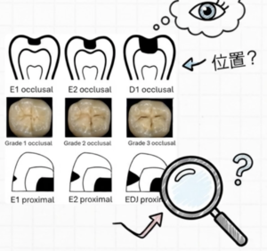
Here, precise assessment based on the international standard ICDAS (International Caries Detection and Assessment System) takes place. For example, the progression of a lesion differs between “ICDAS 1,” which can only be confirmed after drying, and “ICDAS 2,” which is visible even when wet. Identifying these differences at an early stage enables “proactive prevention” that promotes remineralization without drilling.
“Visual examination is recommended as the first-choice method for the detection and assessment of caries on accessible tooth surfaces.” (Source: ORCA-EFCD Consensus Report)
4. [New Common Knowledge 3] “X-rays” are the Golden Key to Uncovering Hidden Risks
While visual-tactile examination is extremely effective, it has limitations in areas where the line of sight cannot reach physically, such as between the teeth (proximal surfaces). The “golden key” here is bitewing radiography. X-rays are performed only when they add value to the results of visual examination, based on the following “intelligent principles”:
- Justification: Clinically judging that the diagnostic benefits obtained from the X-ray certainly outweigh the potential risks of radiation.
- Optimisation: Making every technical effort to obtain the highest quality images with the lowest possible exposure.
| Method | Main Role | Characteristics |
|---|---|---|
| Bitewing | Detecting hidden proximal caries | The modern gold standard. Drastically improves diagnostic accuracy for proximal surfaces. |
| Panoramic | Comprehensive overview of the mouth | Lower resolution than bitewing. Used for surveying the overall structure and bone condition. |
| Dental CT (CBCT) | 3D detailed analysis | Higher radiation dose. Used as an option when precisely investigating the presence of cavitation in complex cases. |
5. [New Common Knowledge 4] A Premium Option: “Radiation-Free”
The latest auxiliary technologies that detect caries without using radiation are extremely valuable options for health-conscious individuals.
- Laser Fluorescence (e.g., DIAGNOdent): Quantifies the depth of a lesion based on the reflection of laser light.
- Near-Infrared Light Transillumination (NILT): Transmits near-infrared light through the tooth to visualize caries as shadows.
These are premium means of providing “peace of mind” for high-risk individuals or pregnant women who require frequent monitoring. However, from a professional standpoint, there are points to note. For example, laser fluorescence carries the risk of “false positives” (showing high numbers even when there is no caries) by reacting to tooth surface staining or biofilm. These are strictly “auxiliary methods” that complement visual inspection and X-rays, and they do not replace the comprehensive judgment of an experienced dentist.
6. [New Common Knowledge 5] AI and Digital Data Engrave the “History of Your Teeth”
With recent digitization, the use of intraoral photography and 3D scanning has evolved from “recording” to “predicting.” Accumulating and comparing data over time has made it possible to objectively grasp the subtle dynamics (monitoring) of lesions.
Automatic diagnostic algorithms using AI (Artificial Intelligence) are also receiving significant attention. However, the cool-headed perspective based on current evidence is “cautious optimism.” According to the ORCA-EFCD report, although AI technology is developing rapidly, there is not yet sufficient verification data for it to become the clinical “gold standard.” It is currently a technology in development, and the fact that the ultimate diagnostic responsibility always lies with the dentist—”human-led medicine”—is the guarantee of trust at this moment.
Conclusion: A Gift to Your Future Self
Utilizing state-of-the-art diagnostic technology to accurately grasp conditions at a “silent stage” before pain occurs is the wisest and most intelligent investment to avoid the risk of future tooth extraction and large-scale restorative treatment.
The next time you sit in a dentist’s chair, try asking your doctor this: “Could you provide me with a detailed assessment regarding the activity and progression stage of my caries?”
This intelligent and proactive question is the key to you enjoying the benefits of cutting-edge preventive dentistry to the fullest and maintaining your own beautiful teeth for a lifetime.
Reference
Kühnisch, J., Aps, J. K., Splieth, C., Lussi, A., Jablonski-Momeni, A., Mendes, F. M., … & Huysmans, M. C. D. (2024). ORCA-EFCD consensus report on clinical recommendation for caries diagnosis. Paper I: caries lesion detection and depth assessment. Clinical oral investigations, 28(4), 227.
Ask question/ Send the Email to Mr. Root canal, Dr. Miyashita
Make an appointment for consultation today.
Tokyo International Dental Clinic Roppongi
- Address: 5-13-25-2nd Floor, Roppongi, Minato-ku, Tokyo
- Phone: 03-5544-8544
- Closest Stations:
- Azabu Juban (Toei Oedo Line take exit7)
- https://youtu.be/iIeG91YEJTA The way to the clinic from Ohedo Line Exit7
- Azabu Juban (Tokyo Metro Namboku Line exit 5a )
- https://youtu.be/3yniFSfucGg The way to the clinic from Namboku Line Exit 5a
- Roppongi (Hibiya Line exit 3)
We look forward to helping you achieve a healthy, beautiful smile!
Hiroshi Miyashita DDS.
Specialist in Periodontology and Endodontics
Certified by the University of Gothenberg, Sweden in 1996