




[AI is Transforming Dental Diagnosis] A Next-Generation Digital Second Opinion That Finds “Hidden Caries” Previously Missed

Caries (cavity) diagnosis
- Introduction: For those who seek certainty in diagnosis Sitting in the dental chair and looking at bitewing radiographs while being told, “There may be a small cavity here. Let’s drill and treat it,” do you truly feel convinced? “Are we over-preparing healthy tooth structure?” “Or missing something instead?” These questions reflect your sincere intent to properly understand and preserve the “asset value” of your teeth.
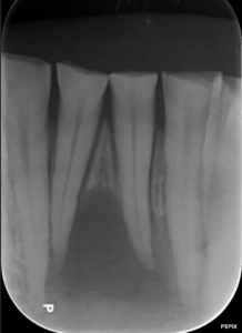
In 2024, a meta-analysis (Ammar & Kühnisch, 2024) regarded as the gold standard in the evidence hierarchy for dental diagnosis was published. Conducted by a research team including Ludwig-Maximilians-Universität München, it provides scientific proof that AI (artificial intelligence) can complement dentists’ vision and dramatically increase diagnostic certainty. In this article, using the latest data, we explore how AI enables a fundamentally minimally invasive approach.
- Point 1: Sensitivity that surpasses the human eye — AI complements the vision of top clinicians A key metric for diagnostic accuracy is sensitivity: the ability to correctly identify sites that truly have caries as “carious.”
According to the latest meta-analysis, AI models showed a pooled sensitivity of 0.87, a high value indicating that AI consistently offers stronger detection performance than traditional dentist-only assessment.
“Compared with dentists, AI models consistently demonstrated a higher mean sensitivity.”
This raises the likelihood of choosing minimally invasive treatment by enabling earlier detection. By passing images through a finely tuned digital filter, AI helps avoid missing subtle initial lesions, making strategic prevention possible to maximize the lifespan of your natural teeth.
- Point 2: The numbers prove it — the striking difference in discriminative power with a DOR of 55.8 When evaluating diagnostic certainty, experts focus on the diagnostic odds ratio (DOR), a single figure that reflects how well a method distinguishes “disease” from “health.”
With conventional dentist visual assessment and radiographs alone, reported DORs typically ranged from 2.6 to 17.11. In contrast, pooled DOR for AI models in the latest research reached 55.8, an exceptionally high value.
This suggests AI can separate “caries” from “healthy tissue” more clearly and with higher confidence. When overwhelming data augments clinical experience, patients’ conviction about the necessity of treatment rises to an unprecedented level.
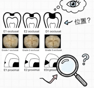
- Point 3: AI’s current status for initial enamel caries To use technology responsibly, we must also be candid about its limitations. For AI as well, diagnostic difficulty varies with lesion depth.
- Dentin caries (deeper lesions): Sensitivity 0.84
- Initial enamel caries (surface lesions): Sensitivity 0.71
Identifying early-stage lesions confined to enamel remains a high-level challenge even for AI. However, pooled specificity (the ability to correctly identify healthy tissue as healthy) is high at 0.89, making AI an excellent advanced assistive tool for flagging suspicious findings.
That said, many AI studies still lack robust external validation (testing in different settings and with different patient data). AI is not a magic wand; it is evolving technology that should be positioned as a partner to the dentist who makes the final clinical judgment.
- Point 4: The importance of specificity to prevent overdiagnosis, and the quality of training data The best care means delivering necessary treatment appropriately while also “protecting teeth that do not need to be drilled.” The concern here is false positives—overdiagnosis that mistakes healthy tissue for disease.
Some studies have found markedly reduced specificity in certain AI models. On analysis, this often traced back to insufficient “true negative” (healthy) images in training, meaning the AI failed to learn “healthy” patterns well.
To avoid “drilling teeth that don’t need drilling,” AI must not only find caries but also reliably recognize healthy tissue. Discerning patients should seek a balanced diagnostic system—one that can confidently declare healthy tissue to be “healthy,” not just point out possible caries.
- Conclusion: A future of dentistry co-created by technology and humans AI does not replace dentists. Grounded in extensive evidence, it strengthens the clinician’s experiential judgment with objective data and elevates diagnostic certainty.
What this meta-analysis shows is an ideal model of dental care: by co-creating with dentists, AI can capture risks that were previously overlooked while simultaneously preventing unnecessary interventions.
Finally, ask yourself: “When entrusting the asset value of my teeth, do I prefer ‘diagnoses backed by data-driven certainty,’ or diagnoses that rely on experience alone?”
The digital transformation of dentistry will serve as a reliable guidepost for greater patient understanding and the pursuit of true oral health.
Reference
Ammar, N., & Kühnisch, J. (2024). Diagnostic performance of artificial intelligence-aided caries detection on bitewing radiographs: a systematic review and meta-analysis. Japanese Dental Science Review, 60, 128-136.
Ask question/ Send the Email to Mr. Root canal, Dr. Miyashita
Make an appointment for consultation today.
Tokyo International Dental Clinic Roppongi
- Address: 5-13-25-2nd Floor, Roppongi, Minato-ku, Tokyo
- Phone: 03-5544-8544
- Closest Stations:
- Azabu Juban (Toei Oedo Line take exit7)
- https://youtu.be/iIeG91YEJTA The way to the clinic from Ohedo Line Exit7
- Azabu Juban (Tokyo Metro Namboku Line exit 5a )
- https://youtu.be/3yniFSfucGg The way to the clinic from Namboku Line Exit 5a
- Roppongi (Hibiya Line exit 3)
We look forward to helping you achieve a healthy, beautiful smile!
Hiroshi Miyashita DDS.
Specialist in Periodontology and Endodontics
Certified by the University of Gothenberg, Sweden in 1996